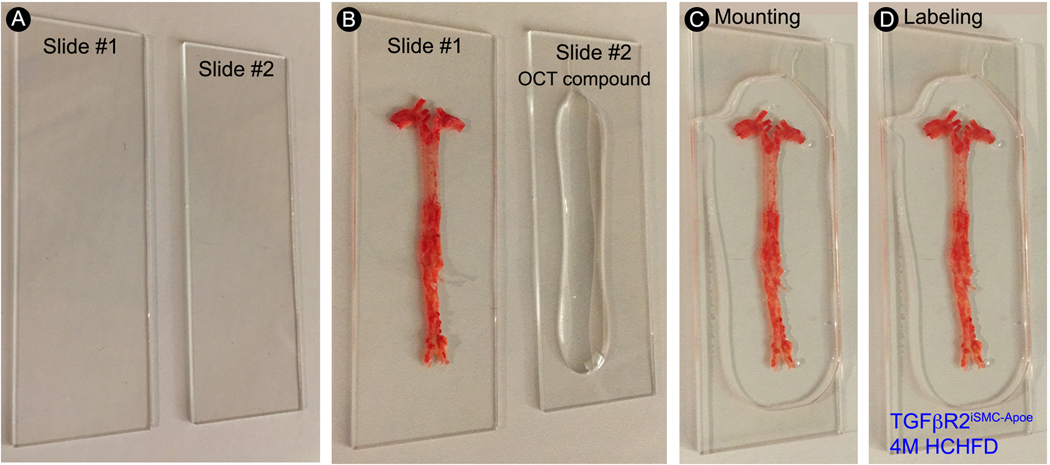
Figure 5.
Step-by-step protocol for en face aorta mounting. (A) Gently clean the glass microscope slides with 70% ethanol and dry with clean laboratory wipes. (B) Apply a small amount of OCT compound to the surface of one glass microscope slide and spread the en face aorta flat on the other glass microscope slide. (C) Gently place the glass microscope slide with OCT compound on the top of the en face aorta sample. (D) Label the slide with sample name.

