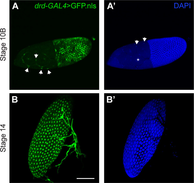
Fig 1. Expression of drd in ovarian follicle cells.

(A, B) GFP expression driven by the drd-GAL4 driver. (A´, B´) DAPI staining of cell nuclei. (A, A´) A stage 10B egg chamber, showing GFP labeling of most follicle cell nuclei. The posterior end of the egg chamber, containing the majority of follicle cells and the oocyte, is to the right, and the anterior end, containing the nurse cells and stretch follicle cells, is to the left. The asterisk in A´ indicates one of the large nurse cell nuclei, which are not labeled with GFP. Arrows in A indicate labeled stretch follicle cell nuclei, and arrow in A´ indicate unlabeled stretch follicle cell nuclei. (B, B´) A stage 14 egg chamber, showing GFP labeling of all follicle cell nuclei. The structures to the right of and overlapping with the egg chamber are respiratory tracheae, which also express drd. Images are maximum intensity projections of Z-stacks. Scale bar is 100 μm.
