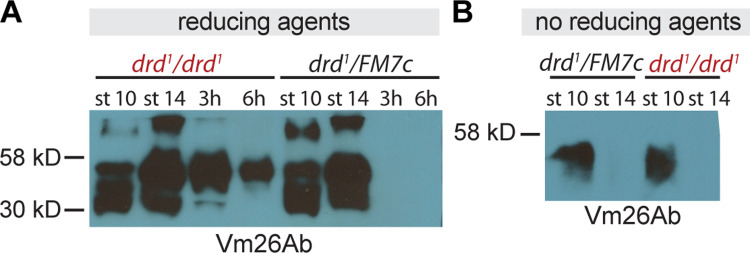
Fig 4. Western blots against vitelline membrane proteins in drd mutant egg chambers and eggs.
(A) Western blot with an antibody raised against Vm26Ab of samples treated with reducing agents. Samples include stage 10 and 14 egg chambers dissected from drd1/drd1 mutant and drd1/FM7c heterozygous females, as well as eggs laid by these females that were collected 0–3 and 0–6 hr after oviposition. 4 eggs or egg chambers per lane, 1:10,000 primary antibody dilution. (B) Western blot from egg chambers solubilized in the absence of reducing agent and probed with an antibody against Vm26Ab. Samples include stage 10 and 14 egg chambers dissected from drd1/drd1 mutant and drd1/FM7c heterozygous females. 2 egg chambers per lane, 1:10,000 Vm26Ab primary antibody dilution.

