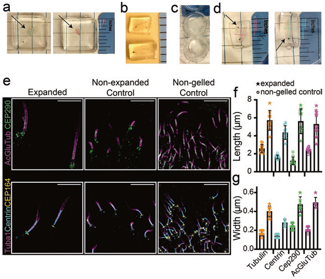
Fig. 1.

Sample preparation during expansion protocol. (a–d) Display examples of the retina in different steps of the protocol, in order: (a) retina immediately after gelation; (b) retina post-disruption, trimmed and slightly expanded; (c) retina after overnight incubation with primary antibodies (opacity subsides once they warm to RT); and (d) orientation of the retinas for embedding in OCT. Arrows point to retinas within the gels, and colored lines display length and width measurements to show that slight expansion of the retinas during disruption and subsequent steps is, by eye, isotropic. Gelled retinas ~0.08 inches wide, ~0.17 inches long; immediately preceding freezing, retinas ~0.13–0.17 inches wide, and ~0.23–0.27 inches long (~0.08 inch changes in each direction). (e) SIM z-projection images displaying cilia, compared in 512 × 512 pixel images, between an expanded section, a non-expanded control section, and a non-gelled control section stained for the antibodies indicated. Scale bar 5 μm. (f, g) Dot plots with averages and standard deviations of the lengths and widths of staining in connecting cilia pre- and post-expansion
