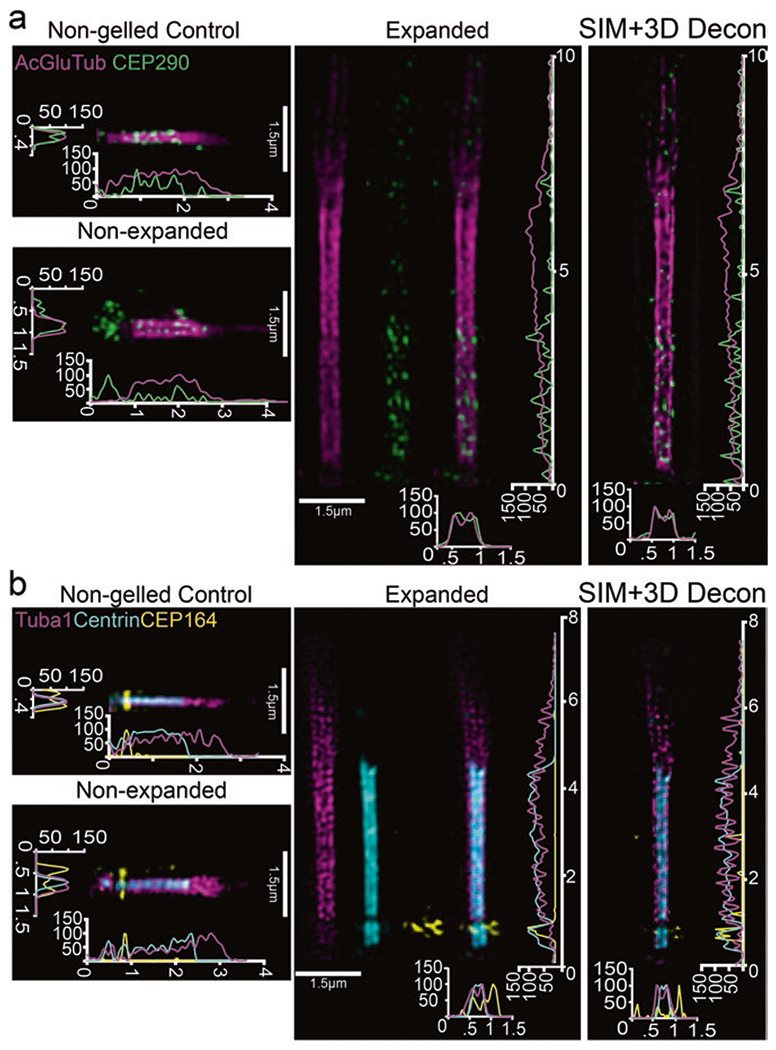
Fig. 2.

Localization of ciliary markers in connecting cilium following expansion in whole retina. (a, b) SIM z-projection images of single, straightened cilia stained for either (a) acetylated alpha-tubulin, glutamylated-tubulin, and CEP290 or (b) tubulin, centrin, and CEP164 with their corresponding controls. SIM z-projections with 3D deconvolution are shown to the right of each panel. On the axes, numbers less than 10 represent μm
