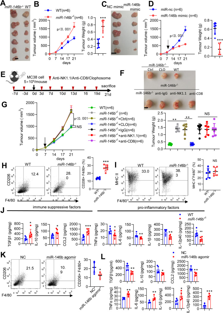
Fig. 1.
miR146b−/− mice exhibit tumor progression in a macrophage-dependent manner. (A-B) WT and miR-146b−/− mice were implanted with 5 × 10⁵ MC38 cells for 3 weeks. Tumor growth and weight were monitored. (C-D) miR-146b−/− mice were implanted with 5 × 10⁵ MC38 cells for 1 week and then treated with miR-nc or miR-146b mimic intraperitoneally twice weekly. Tumor growth and weight were monitored. (E) Mouse models and treatment strategy employed. (F-G) WT or miR-146b−/− mice were pretreated with clodronate and CD8- or NK-depleting antibody every 3 days for 1 week and then injected subcutaneously with MC38 cells. Tumor growth and weight were monitored. n, number of mice. (H-I) Flow cytometry analysis of CD11b+Gr1−F4/80+CD206+ M2-TAMs and CD11b+Gr1−F4/80+MHC II+ M1-TAMs in tumors from WT and miR-146b−/− mice (n= 6). (J) Protein expression of cytokines in tumors from WT and miR-146b−/− mice. (K) Flow cytometry analysis of CD11b+Gr1−F4/80+CD206+ M2 TAMs in tumors from miR-146b−/− mice treated with NC or miR-146b agomir intraperitoneally twice a week (n= 6). (L) Protein expression of cytokines in tumors from miR-146b−/− mice treated with miR-nc or miR-146b mimic

