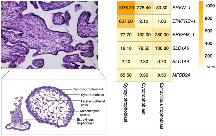
Fig. 2.
Expression levels of selected HERVs and their receptors in placental tissue. Chorionic villi and trophoblast populations in a 28-gestational-week placenta with a schematic cartoon representation of chorionic villus (left). Expression levels of genes coding syncytin-1 (ERVW-1), syncytin-2 (ERVFRD-1), suppressyn (ERVH48-1), and their receptors (SLC1A5, SLC1A4, MFSD2A) in trophoblast populations (right); nTPM, normalized protein-coding transcripts per million. Expression data from proteinatlas.org. The placenta was stained by hematoxylin and eosin. An image of the placenta was taken by light microscope Axio Vert. A1 in software Axio Vision 4.8 (Zeiss) by Lajos Gergely, MD (Institute of Medical Biology, Genetics and Clinical Genetics, Faculty of Medicine, Comenius University Bratislava, Bratislava, Slovak Republic). Created in biorender.com

