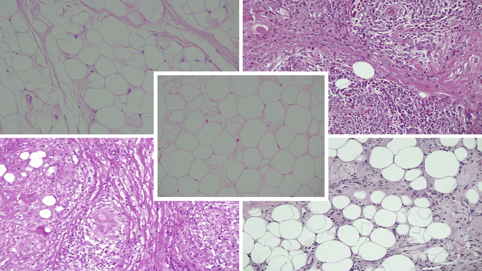
Figure 1.

Specimen grading examples.
Representative images demonstrating the histologic variability of orbital fat in subjects diagnosed with TAO, GPA, sarcoidosis, and NSOI compared to heathy controls. Hematoxylin and eosin staining in paraffin embedded sections (20x).
A. Control (center)
Average inflammation score of 0 (DJW 0, HEG 0). Average fibrosis score of 0.5 (DJW 0, HEG 1).
B. TAO (top left)
49-year-old female within 12 months of ocular onset. No history of steroid or steroid sparing agents or radiotherapy.
Average inflammation score of 0 (DJW 0, HEG 0). Average fibrosis score of 2 (DJW 2, HEG 2).
C. GPA (top right)
24-year-old male within 5 months of ocular onset. No history of steroid or steroid sparing agents or radiotherapy.
Average inflammation score of 3 (DJW 3, HEG 3). Average fibrosis score of 2 (DJW 2, HEG 2).
D. Sarcoidosis (bottom left)
60-year-old female within 8 months of ocular onset. On high dose steroids at time of biopsy without any history of steroid sparing agents or radiotherapy.
Average inflammation score of 3 (DJW 3, HEG 3). Average fibrosis score of 2 (DJW 2, HEG 2).
E. NSOI (bottom right)
83-year-old male within 1 month of ocular onset. On high dose steroids at time of biopsy without any history of steroid sparing agents or radiotherapy.
Average inflammation score of 1 (DJW 1, HEG 1). Average fibrosis score of 1.5 (DJW 1, HEG 2).
