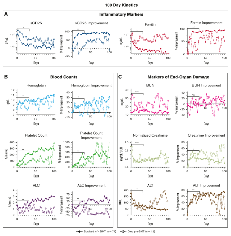
Figure 2.
When followed serially during etoposide-based therapy, multiple HLH-defining and organ-injury markers distinguish patients surviving with HLH from those dying before BMT. Graphs show the serial assessment of the significant absolute laboratory values and their associated improvement from baseline (both plotted as median values) for patients either surviving or dying pre-BMT. Trends in (A) inflammatory markers, (B) blood counts, and (C) markers of end-organ damage were all associated with pre-BMT mortality. Significance was determined using the Mann-Whitney U test assessing values from days 0 to 31 only. Figures show the first 100 days of treatment. Inflammatory markers (A) are displayed on a logarithmic scale. For reference, the day 0 to day 31 median absolute sCD25 was 3864 U/mL for survivors vs 8500 U/mL (overall range, 2939-20 127 U/mL) for patients who died pre-BMT and median absolute ferritin for survivors was 2251 vs 7103 ng/mL (overall range, 1689-17 380 ng/mL) for patients who died pre-BMT. Normalized creatinine was calculated using standard reference ranges for age and gender. Data of nonsignificant absolute laboratory values (P > .05) and their associated improvement from baseline are shown in supplemental Figure 4. Improvement from baseline = (difference between baseline and day X)/baseline value. ∗∗∗∗P < .0001; ∗∗∗P < .001; ∗∗P < .01; ∗P < .05. ALT, alanine transaminase; ns, not significant.

