. 2023 Oct 20;5(6):e230078. doi: 10.1148/rycan.230078

Figure 1:

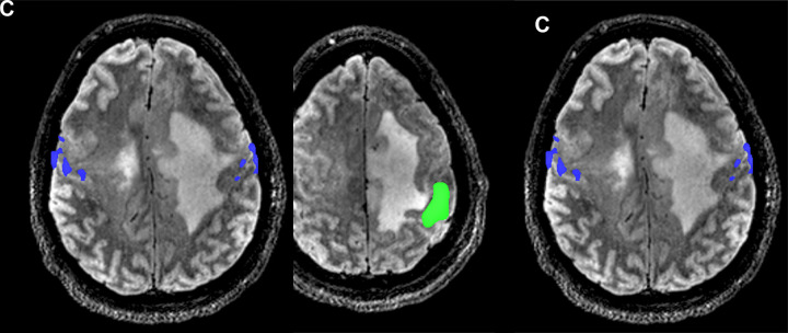
Images from presurgical functional MRI in a 30-year-old man with grade 3 IDH-mutant astrocytoma. (A) Axial image of right foot motor function (magenta) shows localization along the medial aspect of the lesion, with supplementary motor area activation (arrow) in the right hemisphere, suggesting reorganization. (B) Right hand motor function (green) is shown in the left hemisphere hand knob area along the posterolateral border of the lesion. (C) Tongue motor function (blue) is present in the lateral precentral gyrus along the lateral border of the lesion.

Images from presurgical functional MRI in a 30-year-old man with grade 3 IDH-mutant astrocytoma. (A) Axial image of right foot motor function (magenta) shows localization along the medial aspect of the lesion, with supplementary motor area activation (arrow) in the right hemisphere, suggesting reorganization. (B) Right hand motor function (green) is shown in the left hemisphere hand knob area along the posterolateral border of the lesion. (C) Tongue motor function (blue) is present in the lateral precentral gyrus along the lateral border of the lesion.