. 2023 Oct 20;5(6):e230078. doi: 10.1148/rycan.230078

Figure 5:

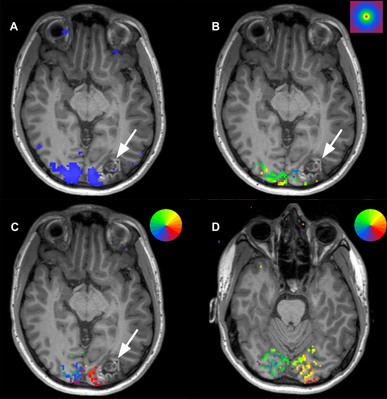
Scans from presurgical retinotopic mapping functional MRI in a 24-year-old man with a left occipital lobe ganglioglioma (arrow). (A) Image obtained during axial checkerboard vision task shows the primary visual cortex (blue) to lie medial to the lesion. (B) Concentric ring paradigm shows the lesion to occupy cortical representation of areas outside of the fovea (stimulus key in right upper corner; fovea = red). (C) Rotating wedge paradigm maps at the level of the lesion show activation in the area of the patient’s inferior outer quadrant, suggesting surgical risk for right inferior quadrantanopia (stimulus key showing color representation of visual fields in right upper corner; the visual field representation in the brain is inverted and flipped right-to-left such that right inferior visual quadrant is represented in the superior left occipital lobe. (D) More inferior image shows that the superior quadrant visual field is not within proximity to the lesion.

Scans from presurgical retinotopic mapping functional MRI in a 24-year-old man with a left occipital lobe ganglioglioma (arrow). (A) Image obtained during axial checkerboard vision task shows the primary visual cortex (blue) to lie medial to the lesion. (B) Concentric ring paradigm shows the lesion to occupy cortical representation of areas outside of the fovea (stimulus key in right upper corner; fovea = red). (C) Rotating wedge paradigm maps at the level of the lesion show activation in the area of the patient’s inferior outer quadrant, suggesting surgical risk for right inferior quadrantanopia (stimulus key showing color representation of visual fields in right upper corner; the visual field representation in the brain is inverted and flipped right-to-left such that right inferior visual quadrant is represented in the superior left occipital lobe. (D) More inferior image shows that the superior quadrant visual field is not within proximity to the lesion.