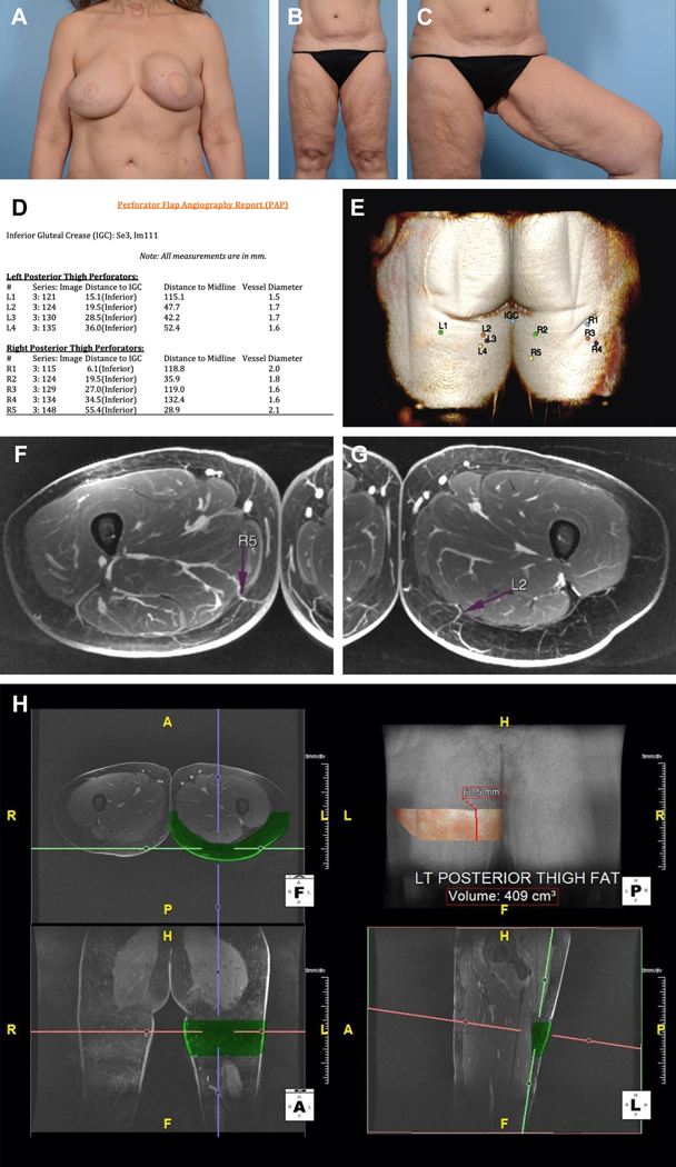
Figure 1.
Preoperative images of the (A) breasts and (B, C) lower extremities. Patient had a history of right breast cancer and underwent mastectomy with DIEP flap reconstruction. The left breast subsequently developed cancer and was reconstructed with a pedicled latissimus dorsi flap and implant. Note severe capsular contracture of left breast a well as laxity of tissue in the posteromedial thighs. Magnetic resonance angiography (MRA) of the bilateral lower extremities with and without intravenous contrast and 3-dimensional (3-D) postprocessing was performed. (D) Perforator flap angiography report detailing vessel caliber as well as location. IGC=Inferior Gluteal Crease. (E) 3-D perforator map showing perforator location on posterior thigh. (F) Axial view of the thigh demonstrating a favorable R5 perforator located 55.4 mm inferior to the IGC, 28.9 mm to the right of midline, and 6.9 mm posterior to the posterior margin of gracilis. Vessel diameter is 2.1 mm. It travels 185.2 mm with an intramuscular course before joining the inferior gluteal artery. (G) Axial view of the thigh demonstrating a favorable L2 perforator located 19.5 mm inferior to the IGC, 47.7 mm to the left of midline, and 39.5 mm posterior to the posterior margin of gracilis. Vessel diameter is 1.7 mm. It travels 185.2 mm with an intramuscular course before joining the inferior gluteal artery. (H) Fat volume of a 22×6 cm flap on posterior left thigh is 409.0cc

