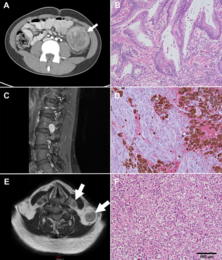
FIGURE 3.
Imaging and histology for 3 highlighted patients. A, Axial CT image of the abdomen showing a mass in the descending colon (arrow) in a 15-year-old male diagnosed with poorly differentiated adenocarcinoma. B, Histology shows high dysplasia and stromal microinvasion. C, MRI of the lumbar spine in a 36-year-old female diagnosed with malignant melanotic schwannoma shows oval-shaped mass in left neural foramen of L3-L4 with avid enhancement on sagittal T1-weighted fat-suppressed image (arrow). Histology (D) shows pigmented spindle and epithelioid cells. E, MRI displays two left supraclavicular masses on T1-weighted image in a 75-year-old male with metastatic poorly differentiated carcinoma of unknown primary in the neck. F, Histology shows epithelioid tumor cells. Scale bar = 100 µm and is the same for B, D, and F.

