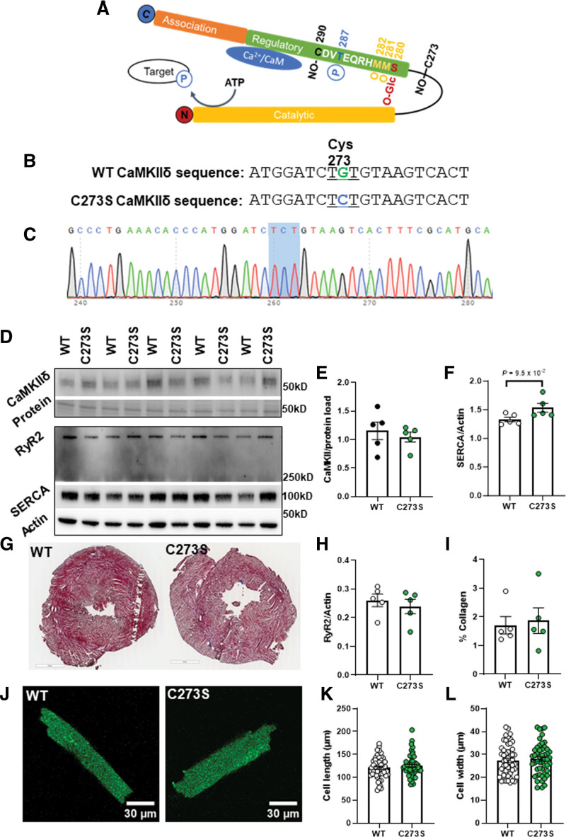
Figure 3.
Characterization of the CaMKIIδ (Ca2+/calmodulin kinase II delta)-C273S knock-in mouse model. Schematic of the CaMKIIδ monomer with the residue positions of various posttranslational modifications within the regulatory domain including S-nitrosylation (NO-), phosphorylation (P), oxidation (O), and O-GlcNAc modification, which are known to alter CaMKII activity (A). A mouse model was generated using CRISPR/cas9 causing a single point mutation in the cystine codon at position 273, resulting in replacement with a serine residue which cannot be S-nitrosylated (B). Example of a CaMKIIδ-C273S offspring confirmed with genotyping of ear notch (C). Protein expression in ventricular tissue (N=5 hearts) was measured using Western blots (D) for CaMKIIδ (E), SERCA (SR Ca2+ ATPase; F), RyR (ryanodine receptor type; H), and normalized to actin or protein load. Ventricular fibrosis was measured in fixed and stained tissue (G) by quantifying collagen content (I). Representative cardiomyocytes are shown in J for measurement of cell length (K) and width (L), wild type (WT): n=52, N=7 hearts; CaMKIIδ-C273S: n=50, N=8 hearts.

