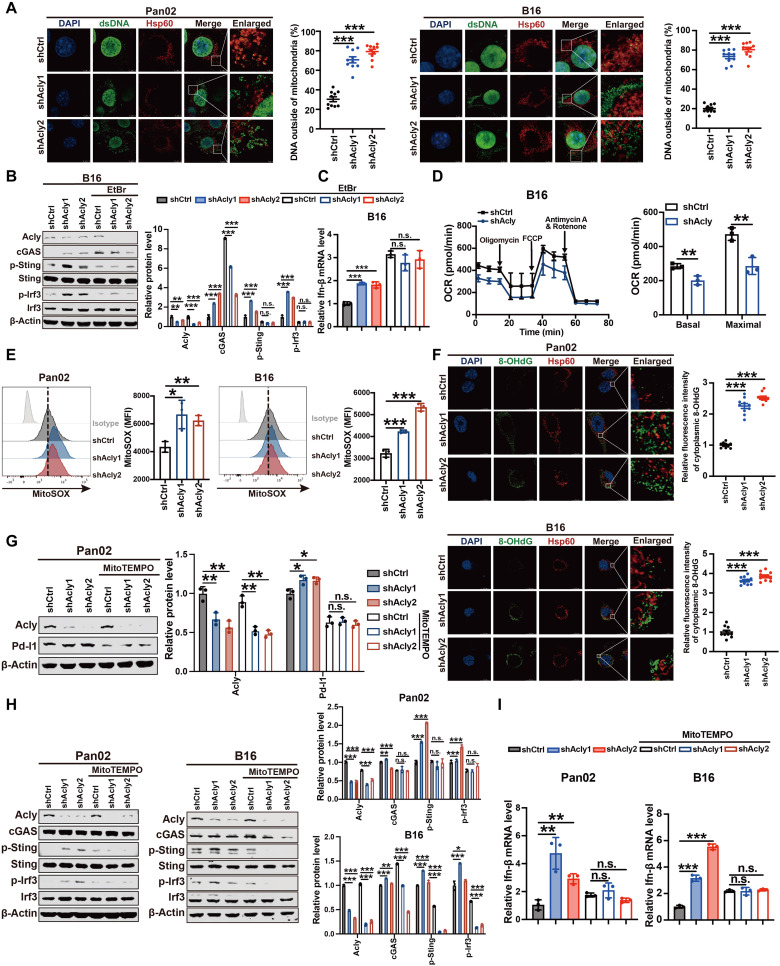
Fig. 3. ACLY inhibition–induced mitochondrial damage triggers mtDNA leakage to activate cGAS-STING signaling.
(A) Immunofluorescence costaining of anti-dsDNA (green), anti-Hsp60 (red), and 4′,6-diamidino-2-phenylindole (DAPI) (blue) in Pan02 cells and B16 cells with Acly knockdown or not. The percentage of dsDNA (green) outside of mitochondria (red) was quantified. Scale bars, 5 μm. (B) Immunoblot analysis of indicated protein expressions in B16 cells with Acly knockdown or not after EtBr treatment. (C) qPCR analysis of Ifn-β mRNA expression in B16 cells with Acly knockdown or not after EtBr treatment. (D) Seahorse analysis of oxygen consumption rate (OCR) and maximal respiration in B16 cells with Acly knockdown or not. (E) Flow staining and MFI of mitoSOX in Pan02 cells and B16 cells with Acly knockdown. (F) Immunofluorescence costaining of anti-DNA damage (8-OHdG) (green), anti-Hsp60 (red), and DAPI (blue) in Pan02 cells and B16 cells with Acly knockdown or not. The relative fluorescence intensity of cytoplasmic 8-OHdG (green) outside of mitochondria (red) was quantified. Scale bars, 11.5 μm. (G) Immunoblot analysis of indicated protein expressions in Pan02 cells with Acly knockdown or not after mitoTEMPO treatment (10 μM). (H) Immunoblot analysis of indicated protein expressions in Pan02 cells and B16 cells with Acly knockdown or not after mitoTEMPO treatment (10 μM). (I) qPCR analysis of Ifn-β mRNA expression in Pan02 cells and B16 cells with Acly knockdown or not after mitoTEMPO treatment (10 μM). n = 10 biological replicates from two independent experiments [(A) and (F)] and data are shown as means ± SEM; n = 3 biological replicates from three independent experiments [(B), (G), and (H)]; n = 3 biological replicates from one independent experiment [(C) to (E) and (I)]. Statistical significance was assessed by one-way ANOVA [(A) to (C) and (E) to (I)] and unpaired t test (D); *P < 0.05; **P < 0.01; ***P < 0.001.

