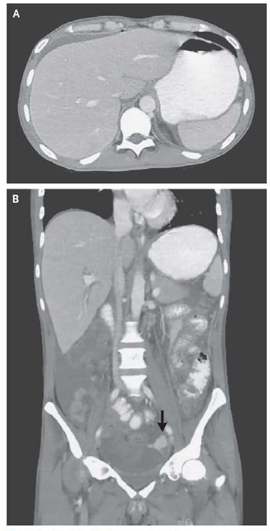
Figure 3: CT of the Abdomen and Pelvis.

CT of the abdomen and pelvis was performed after the adminstraion of oral contrast material. An axial iamge of the abdomen (Panel A) shows a punctate focus of pneumoperitoneum anterior to the liver, with small volume perihepatic ascites. A coral image of the abdomen and pelvis (Panel B) shows diffuse wall thickening in the rectosigmoid colon and extravasation of extraluminal contrast material (arrow) into the area adjucent to the sigmoid colon, with layering of the contrast material, findings that are thought to indicate a perforation.
