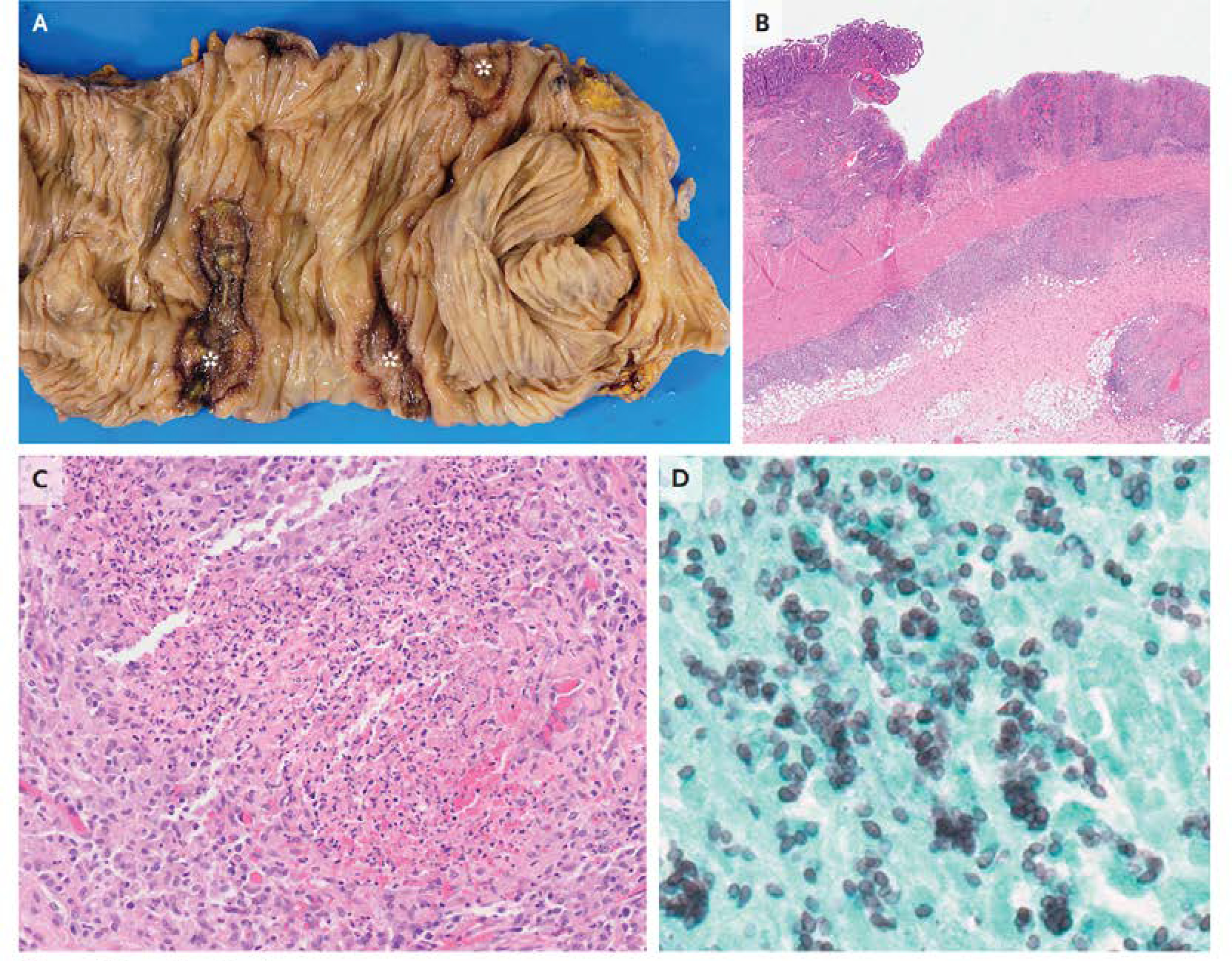
Figure 4: Resection Specimens.

Gross examination of resection specimen of the colon (Panel A) reveals ulceration (asterisks). Hematoxylin and eosin staining (Panel B) shows ulceration with transmural granuloma. At higher power (Panel C), necrotizing granulomas are seen. Grocott-Gomori methenamine silver staining (Panel D) highlights clusters of small, ovoid yeast forms that are consistent with Histoplasma capsulatum.
