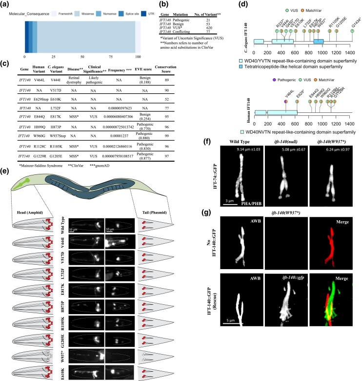
Fig. 2.
The ift-140W937stop mutants phenocopy defects of loss-of function ift-140 mutants. a) Shown are the percentage distribution of the variation types for human IFT140 in the ClinVar. b) Number of missense variants in human IFT140 from ClinVar is displayed with clinical significance. c) Caenorhabditis elegans variants in IFT-140 and human variants in IFT140 corresponding to the human positions of C. elegans variants are presented. Clinical significance, disease and frequency (downloaded from gnomAD), EVE score, and conservation score (see Materials and Methods) were presented for human variants. d) The variants, including matching variants (MatchVars) from humans and C. elegans IFT140 were displayed in their corresponding positions. VUS means variant of uncertain significance. e) Fluorescence images from the head and tail were displayed with representative drawings following the dye-filling assay. C. elegans cartoon shows the whole worm. The intermittent line in fluorescence images marks the edge of worms in the head and tail. With exception of mutants carrying W937stop, wild type and other worms absorb the lipophilic fluorescent dye in the head and tail. Asterisk (*) refers to the stop codon. Scale bar: 10 μm. f) Shown are the localization of IFT-74::GFP in the tail cilia (phasmid) of wild type, ift-140(lf), and ift-140W937stop. Scale bar of 3 μm. The average tail cilia lengths for wild type, ift-140(lf), and ift-140W937stop were presented. g) Shown are images depicting the AWB cilia in ift-140(W937*) mutants and the rescued line. The rescued line consists of ift-140(W937*) mutants expressing extrachromosomal IFT-140::GFP.

