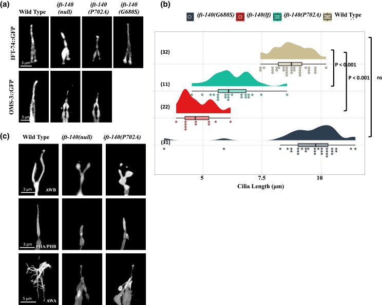
Fig. 4.
The sensory cilia in the C. elegans IFT-140P702A mutants are shortened. a) Shown are confocal images displaying the localization of fluorescently tagged IFT proteins in the tail (phasmid) of wild-type and the indicated mutants. Scale bar: 3 μm. b) Plots of the PHA/PHB cilia length for wild-type and indicated mutants were shown. Not significant is abbreviated as ns. P values between the designated mutants and the wild type are also presented. Numbers in parentheses indicate the number of cilia used to calculate cilia length. c) Fluorescent markers display the following cilia: AWB cilia (Y-shaped cilia), PHA/PHB cilia (rod-shaped cilia), and AWA cilia (multiple complex branches). Shown are representative fluorescent images from wild type, ift-140(lf), and ift-140P702A mutants. Scale bar (AWB): 3 μm, scale bar (PHA/PHB): 3 μm, and scale bar (AWA): 5 μm.

