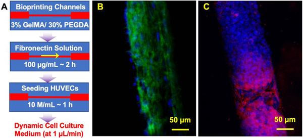
Figure 5. Vascular modeling in 3D bioprinted microfluidic chip:
(A) The procedure to seed HUVECs and put dynamic flow; B) fluorescence images showing HUVECs stained for F-actin (green) and nuclei (blue) at Day 10; and C) fluorescence images showing HUVECs stained for CD31 (red) and nuclei (blue) at Day 10.

