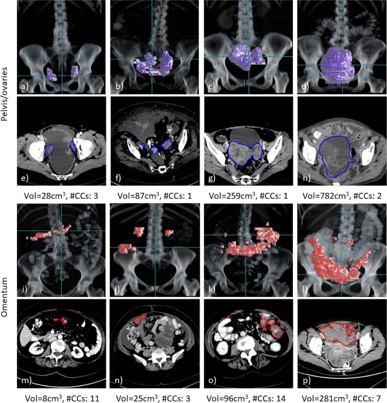
Fig. 1.
Examples of three-dimensional volume renderings (a–d, i–l) and axial slices (e–h, m–p) for pelvic/ovarian and omental lesions of high-grade serous ovarian carcinoma patients. For each example, the ground truth tumour volume (Vol) and number of connected components (#CCs) are shown. The scans shown were all contained in the training set and selected such that their lesion volume equals the 25, 50, 75, and 90 percentiles of the lesion volume in the training set (left to right). The horizontal green line in the rendering visualisations corresponds to the axial slice shown below. Both disease sites demonstrate a great variability of disease expression among different patients, which poses a great challenge for manual and automated segmentation methods

