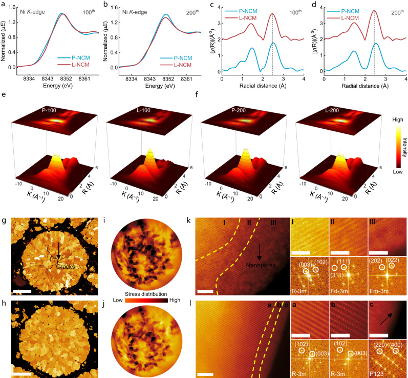
Fig. 5. Coordination environment, morphology, and microstructure investigations.
a, b Ni-K edge XANES spectra for 100th (a) and 200th (b) cycled electrodes. c, d FT-EXAFS spectra for 100th (c) and 200th (d) cycled electrodes. e, f Wavelet-transformed EXAFS for 100th (e) and 200th (f) cycled electrodes. g, h Cross-section SEM images for P-NCM (g) and L-NCM (h) after 200 cycles. Scale bars, 5 μm. i, j Stress distributions for P-NCM (i) and L-NCM (j) after 200 cycles (the mode based on the cross-section SEM image). k, l HAADF-STEM and FFT profiles for P-NCM (k) and L-NCM (l) after 200 cycles. Left, scale bars, 5 nm; right (enlarged area), scale bars, 2 nm.

