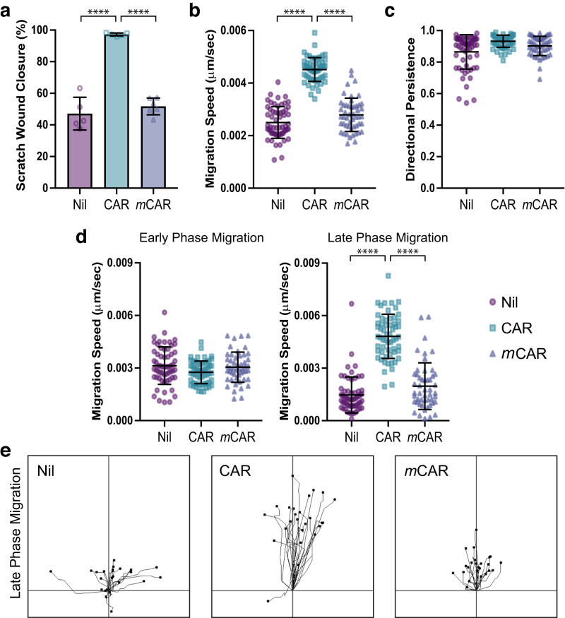
Fig. 3. CAR peptide stimulates keratinocyte migration.
Migration of HaCaT keratinocytes on fibronectin in scratch wound assays, in the presence or absence of 10 µg/ml CAR or mCAR peptide. Cells were analysed over 20 h by time-lapse microscopy. a Scratch wound closure, b mean migration speed throughout timelapse, c directional persistence throughout timelapse, d speed at early and late phases of migration (Early: Timepoint 0–5 h; Late: Timepoint 15–20 h), and e representative migration tracks during late phase migration. Data are representative from one of three independent experiments. Values are means ± S.D. All statistical analyses are two-way ANOVA with Tukey’s multiple comparisons test. a n = 5–6 fields of view per condition (Nil n = 5, CAR n = 5, mCAR n = 6); Nil vs CAR P = 6.133 × 10⁻⁸; CAR vs mCAR P = 1.147 × 10⁻⁷. b–d n = 50–60 cells per condition (Nil n = 60, CAR n = 59, mCAR n = 50). b Nil vs CAR P = 1.5 × 10⁻¹⁴; CAR vs mCAR P = 1.5 × 10⁻¹⁴. d Early phase: Nil vs CAR P = 0.0499; CAR vs mCAR P = 0.1932. Late phase: Nil vs CAR P = 1.5 × 10⁻¹⁴; CAR vs mCAR P = 1.7 × 10⁻¹⁴. a Each data point represents a single field of view; b–d Each data point represents an individual cell. Source data are provided as a Source Data file. See also Supplementary Movies S1 & S2.

