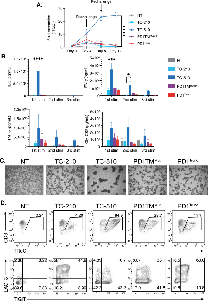
Fig. 4.
Chimeric PD-1 receptor confers enhanced fitness to TRuC-T cells during a repeated stimulation assay. a Fold expansion of TRuC-T cells normalized for transduction efficiency and cultured with MSTOMSLN-PD-L1 tumor cells at a 1:20 effector-to-target ratio. b MSD ELISA results from culture supernatants collected 72 h after each antigen challenge and analyzed for cytokines. c Brightfield microscopy images showing visible clustering in cultures containing TC-510 T cells at Day 8. d FACs plots of CD3 and TRuC receptor expression on viable CD45+ from MSTOMSLN-PD-L1 cultures on Day 12 of culture. Statistical analysis was carried out with a two-way ANOVA. *p < 0.05, ***p < 0.01, ****p < 0.0001. Data are representative of three independent donors and are plotted as mean (± SEM). ELISA enzyme-linked immunosorbent assay, GM-CSF granulocyte macrophage colony-stimulating factor, IFN-γ interferon gamma, IL interleukin, LAG-3 lymphocyte activation gene 3, MSD meso scale discovery, MSLN mesothelin, MSTO mesothelioma, Mut mutant, NT nontransduced, PD-1 programmed cell death protein 1, PD-L1 programmed cell death protein ligand 1, SEM standard error of the mean, TIGIT T cell immunoglobulin and ITIM domain, TNF-α tumor necrosis alpha, TRuC T cell receptor fusion construct

