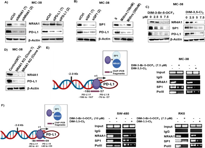
Fig. 2.
Expression of PD-L1 in mouse MC-38 colon cancer cells and regulation by NR4A1. MC-38 cells were transfected with oligonucleotides targeting NR4A1 and PD-L1 (A), Sp1 or treated with mithramycin (B) or treated with DIM-3-Br-5-OCF3 and DIM-3,5-Cl2 (C) or stable MC-38 NR4A1-KO cells generated by CRISPR/Cas9 and two clones (Clone 7 and Clone 14) (D), and whole cell lysates were analyzed by western blots. E Model of the GC-rich mouse PD-L1 promoter and primers targeting this region for the ChIP assay. MC-38 cells were treated with DMSO, 7.5 µM DIM-3-Br-5-OCF3 and 7.5 µM DIM-3,5-Cl2 and interaction with the GC-rich PD-L1 gene promoter was determined in a ChIP assay. F Model of the GC-rich human PD-L1 promoter and primers targeting this region in the ChIP assay; SW480 and RKO cells were treated as described and interactions with the PD-L1 promoter were determined in a ChIP assay as outlined in Methods. Figures were created partly with BioRender (https://app.biorender.com)

