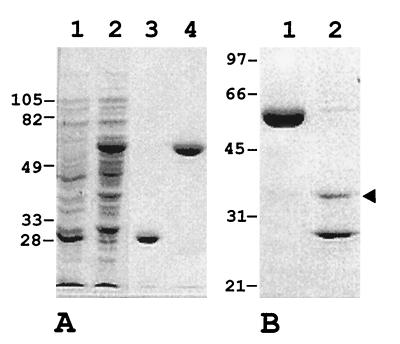
FIG. 3.
Expression in E. coli and purification of a GST-MoaC fusion protein. (A) SDS-PAGE of a cell extract from strain BL21(pGEX-4T-2) containing GST (lane 1), cell extract from BL21(pCSLM43) containing GST-MoaC (lane 2), purified GST (lane 3), and purified GST-MoaC protein (lane 4). (B) SDS-PAGE of purified GST-MoaC protein (lane 1) and thrombin-treated GST-MoaC protein (lane 2). The arrowhead points to the ca. 36-kDa Synechococcus MoaC protein. Positions and sizes (in kilodaltons) of some molecular weight markers are indicated to the left of each panel.

