
Figure 1.
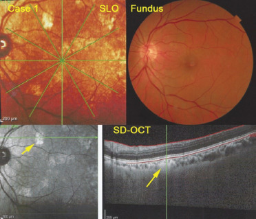
Multimodal imaging of Case 1. SLO of the left eye (top left) showing whitish hyperreflective lesions characteristic for melanocytic lesions, while on classical fundus photographs of the posterior pole hyperpigmented lesions are barely detectable (fundus, top right). On OCT, lesions appear as fluffy hyperreflective lesions just underneath the RPE-Bruch's membrane complex (yellow arrow on SD-OCT) and correspond to whitish areas on infrared frame (bottom left, yellow arrow). SLO: Scanning laser ophthalmoscopy