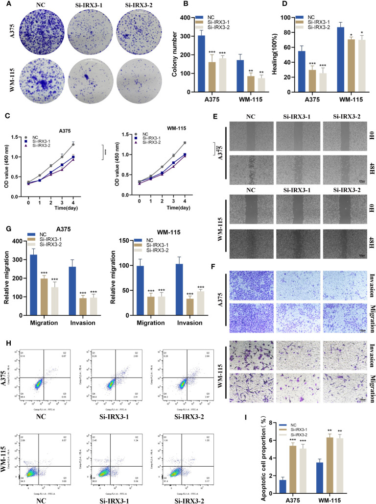
Figure 10.
Silencing IRX3 Inhibits Proliferation, Invasion, and Metastasis while Promoting Apoptosis in Melanoma Cells. (A) A colony formation assay was performed on A375 and WM-115 melanoma cells in the NC and si-IRX3 groups. Smaller colonies were observed in the si-IRX3 group, indicating that IRX3 silencing inhibits melanoma cell proliferation. (B) Quantification of colony formation assay results showed a decrease in colony size in the si-IRX3 group compared to the NC group. (C) CCK-8 assay further confirmed the inhibitory effect of IRX3 silencing on melanoma cell proliferation. (D) Quantification of scratch assay results showing a decrease in wound closure percentage in the si-IRX3 group compared to the NC group. (E) Scratch assay revealed a decrease in 17 the migration ability of A375 and WM-115 cell in the si-IRX3 group compared to the NC group. (F) Transwell assay demonstrated a decrease in the invasion ability of A375 and WM-115 cells in the si-IRX3 group compared to the NC group. (G) Quantification of transwell assay results shows a decrease in the number of invading cells in the si-IRX3 group compared to the NC group. (H) Apoptosis assay revealed an increase in apoptosis in both A375 and WM-115 cell lines upon IRX3 silencing. (I) Quantification of apoptosis assay results shows an increase in the percentage of apoptotic cells in the si-IRX3 group compared to the NC group. *P < 0.05, **P < 0.01, ***P < 0.001.

