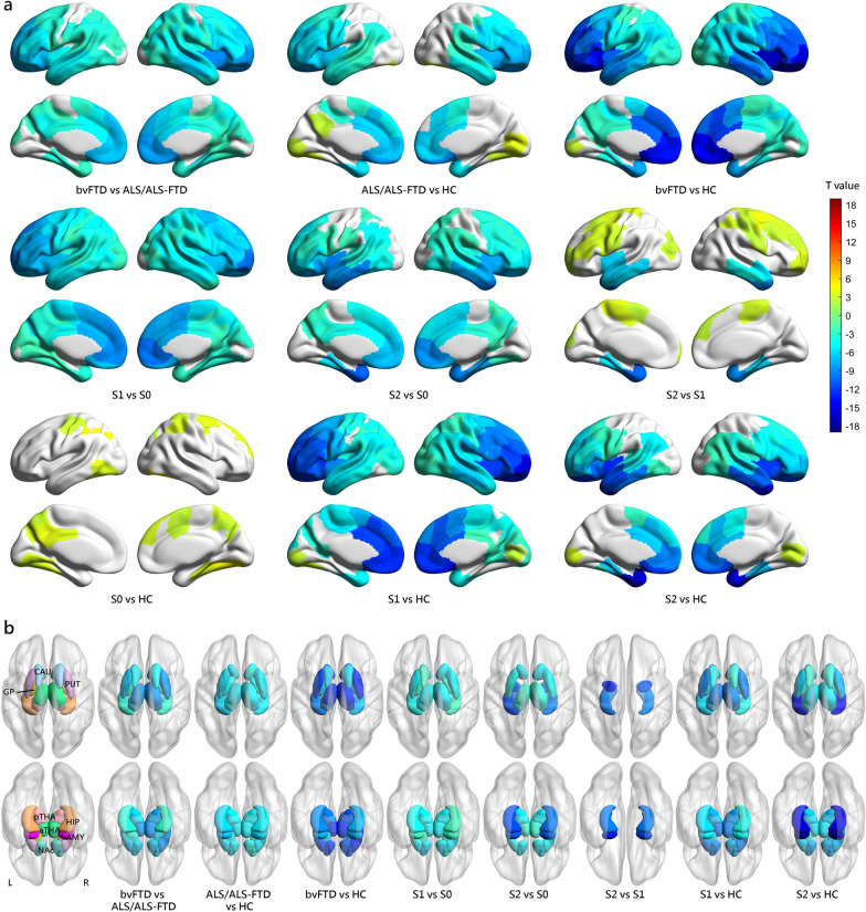
Fig. 5.
Comparison of volumetric measurements between groups at baseline. a Cortical volumetric differences between groups at baseline. b Subcortical volumetric differences between groups at baseline. Only results with a threshold at FDR-corrected P < 0.05 are shown. Cool colors indicate more cortical atrophy in the former group than the latter one, while warm colors indicate more cortical atrophy in the latter group than the former one. S0 Normal-appearing group, S1 Prefrontal/Somatomotor-predominant subtype, S2 Limbic-predominant subtype

