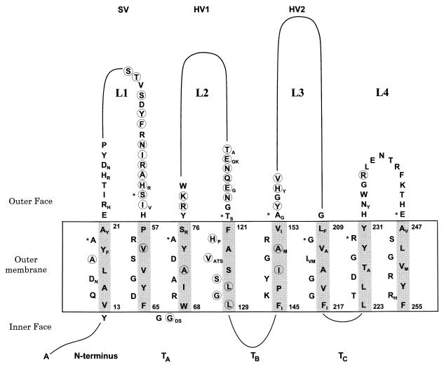
FIG. 4.
Predicted two-dimensional structure of Opa proteins. Variable stretches are indicated by a continuous line, and only details of conserved amino acids are shown. Minor sequence variation is indicated by additional subscript letters next to the most frequent amino acids. Circled amino acids were variable only in the Opa proteins from N. flava or N. sicca. Asterisks indicate the first amino acids predicted to lie outside the outer membrane in the model of Bhat et al. (4). The inner and outer faces of the outer membrane are indicated by horizontal lines. The gray shading indicates the nonpolar side of the eight transmembrane β strands.

