
Figure 5. Accumulation of GNL3 into the nucleolus limits origin firing.
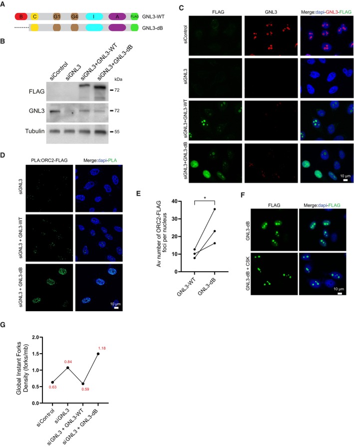
- Schematic representation of human GNL3 protein with its associated domains (B: basic domain; C: coiled‐coil domain; G1: GTP‐binding motif 1; G4: GTP‐binding motif 4; I: intermediate domain; A: acidic domain). GNL3‐WT and GNL3‐dB are fused with FLAG.
- Western‐blot analysis of Flp‐In T‐Rex HeLa cells expressing exogenous GNL3‐WT or GNL3‐dB. Cells were transfected with siControl or siGNL3 for 48 h then expression of exogenous GNL3‐FLAG (resistant to the siRNA against GNL3) was induced using 10 μg/ml of doxycycline for 16 h.
- Immunofluorescence analysis of Flp‐in T‐Rex HeLa cells expressing exogenous GNL3‐WT or GNL3‐dB.
- PLA (proximity ligation assay) analyzing the proximity between ORC2 and GNL3‐FLAG or GNL3‐dB‐FLAG in HeLa Flp‐In cells upon doxycycline induction.
- Graphic representation of the average number of PLA ORC2‐FLAG foci in three biological replicates. For statistical analysis paired t‐test was used; *P < 0.05.
- Immunofluorescence experiment of HeLa Flp‐In cells expressing GNL3‐dB with or without pre‐extraction with cytoskeletal buffer (CSK).
- Analysis of GIFD (Global Instant Fork Density) by DNA combing in HeLa cells (n = 1; a biological replicate is shown in Fig EV5C). GIFD value is indicated in red.
Source data are available online for this figure.