
Figure EV1. Generating OTCs.
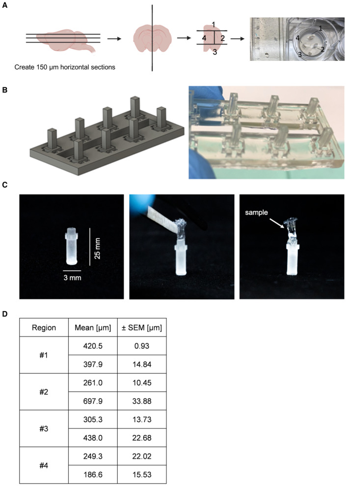
- Schematic setup of generating 150‐μm‐thick mouse brain slices (OTC) and cutting them along the hemisphere into four intersections and placing them on a Millicell culture insert. Scheme created with BioRender.com.
- 3D rendering of the CAD design of the thermoforming positive molds for the LSFM holders. An array of eight positive molds is shown. The molds have a square 2 × 2 mm cross‐section and a height of 5 mm. On the right panel, the real 3D‐printed molds array is displayed.
- One thermoformed FEP‐foil LSFM holder (3 × 25 mm) glued to a sample holder, followed by an OTC is placed inside (right panel).
- Table depicting the mean and SEM of the tumor cell dispersion of one biological replicate (n = 2 tumors per region) of spheres placed onto different brain regions depicted in (A) using GraphPad Prism 9.