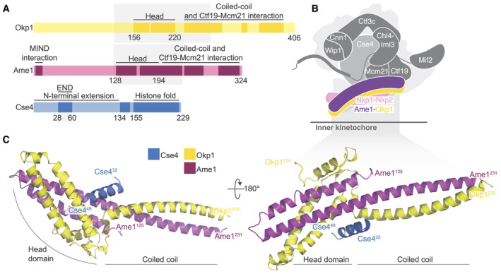
Figure 1. Crystal structure of the Okp1‐Ame1‐Cse4END complex.

- Domain diagram showing the relevant regions of Okp1, Ame1, and Cse4. The shaded boxes demarcate the minimal Okp1‐Ame1 peptides used for crystallography. The “Head” domains correspond to the previously described four‐helix bundle (by analogy to the MIND complex; Dimitrova et al, 2016). Darker regions of the primary structure diagram indicate segments that are ordered in previously reported structures or that are known to bind partners. An N‐terminal peptide of Ame1 connects to spindle microtubules indirectly via the MIND complex (Hornung et al, 2014).
- Schematic view of the yeast inner kinetochore (Ctf19c, Mif2, and Cse4) showing the position of Okp1‐Ame1 (purple and yellow) and Nkp1‐Nkp2 (pink). N‐terminal extensions are omitted for clarity.
- Crystal structure of the Okp1‐Ame1‐Cse4END complex (this work). Protein chains are colored as in panel A.
Source data are available online for this figure.
