
Figure EV3. Multichannel microscopy images of BrdU‐S100β‐Sox2 immunofluorescence (related to Fig 5).
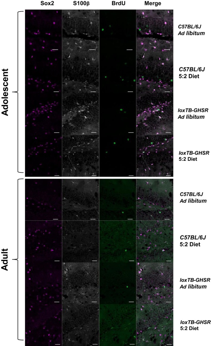
Images were acquired with an LSM980‐Airyscan2 confocal system (Zeiss), using the SR‐4Y airyscan mode. Scale bars represent 20 μm. BrdU = 488 nm; S100β = 568 nm; Sox2 = 647 nm.
Source data are available online for this figure.