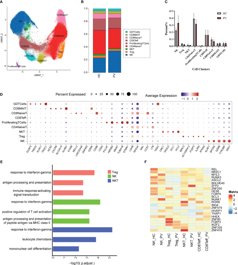
Figure 2.
The heterogeneity and transcriptional features of NK&T cells in PV patients. (A) UMAP representation of 91,224 NK&T cells, showing the formation of nine clusters. (B) The fraction of cells for nine types in HCs and PV patients. (C) Bar graphs of each cell cluster population between HCs and PV patients. (D) Bubble plot of top5 gene expression in each cell cluster; the size of bubble represents the percentage of expressed cells; the color represents the average expression of each gene in clusters: red means the high expression. (E) GO analysis showing the biological process enriched in Treg NK and NKT of PV patients. (F) Heatmap showing the average expression of key regulatory TFs (estimated using SCENIC) between NK cells and T cell subsets in PV patients and HCs.

