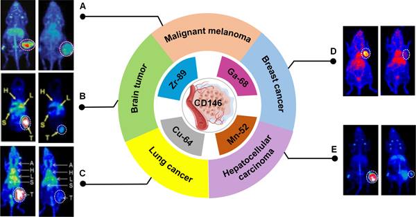
Figure 2. CD146-targeted imaging via nuclear medicine.
(A) 89Zr-Df-YY146 (left) and 89Zr-Df-IgG PET (right) imaging enable visualize CD146-expressing A375 xenografts clearly[69]. (B) PET images of 64Cu-NOTA-YY146 in mice containing U87MG (left) and those are preinjected with a blocking dose of unlabeled antibody (right)[27]. (C)PET Images of tumor-bearing mice derived from lung cancer. H460 (left) and H522 (right) tumors have shown the highest and lowest tracer accumulation, respectively[70]. (D) 64Cu-NOTA-YY146 PET imaging in MDA-MB-435 (left) and pre-blocking (right) tumor models[35]. (E) PET images of mice bearing HepG2 and mice pre-injected with a blocking dose of YY146[71]. There is a white dashed circle indicating the tumor’s location.

