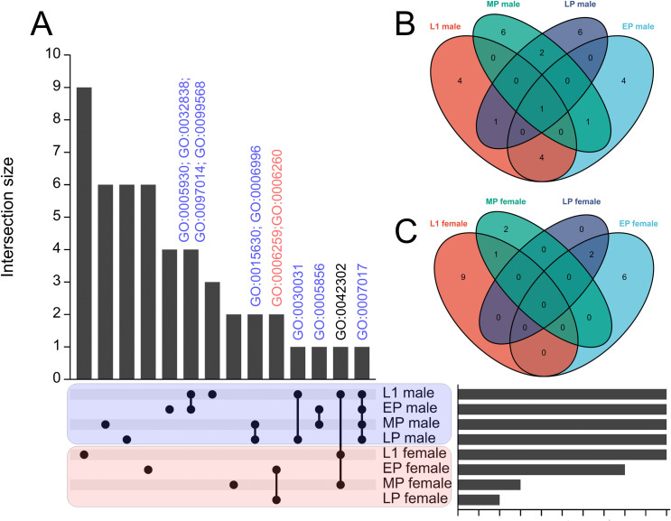
Fig 3. Comparing the transcriptome across larvae and pupae stages of mosquito.
SEPARATOR mosquitoes were utilized to individually segregate male and female mosquitoes at the L1 larval stage using the GFP signal. Following separation, total RNA extraction and RNA-seq analysis were performed. The analysis of the early pupae (EP), mid pupae (MP), and late pupae (LP) stages was conducted using data from a previous study. Sexing at the pupae stages relied on sex-specific morphological differences. Sex-enriched genes were identified using DESeq2 and then performed GO enrichment analysis. The shared genes between the L1 stage and all other comparisons were determined, and visualizations such as (A) UpSet plots and (B-C) Venn diagrams were created to represent these shared genes. The common GO terms across various developmental stages of mosquitoes are labeled above the bar. GO:0005930 axoneme; GO:0032838 plasma membrane bounded cell projection cytoplasm; GO:0097014 ciliary plasm; GO:0099568 cytoplasmic region; GO:0015630 microtubule cytoskeleton; GO:0006996 organelle organization; GO:0006259 DNA metabolic process; GO:0006260 DNA replication; GO:0030031 cell projection assembly; GO:0005856 cytoskeleton; GO:0042302 structural constituent of cuticle; GO:0007017 microtubule-based process.

