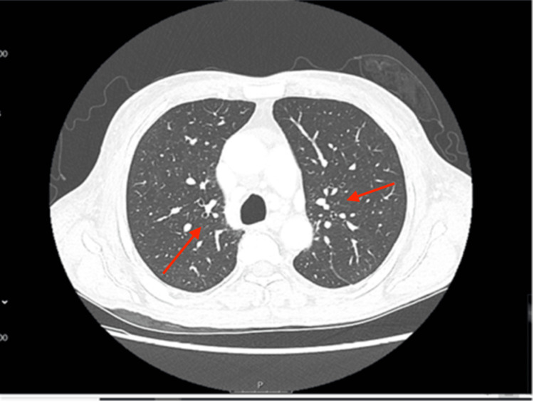
Figure 2. CT thorax with contrast showing interstitial pneumonia pattern of interstitial lung disease (red arrow).
The image revealed no pulmonary nodules, masses, or focal airspace consolidations. However, the underlying pulmonary infectious process could not be excluded. In addition, the image revealed cardiomegaly with pericardial effusion and mediastinal lymphadenopathy.
CT: computed tomography.

