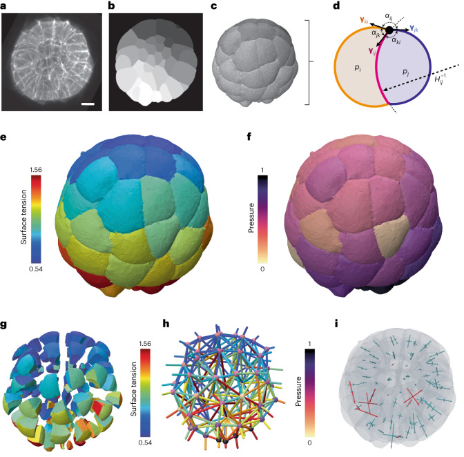
Fig. 2. 3D force-inference procedure and resulting mechanical atlas for a 64-cell ascidian embryo.
a, 3D fluorescence microscopy image (maximum projection) of a 64-cell P. mammillata embryo (from ref. 19). Scale bar, 20 μm. b, Cell segmentation mask in one focal plane of the 3D image. c, Multicellular surface mesh of cell interfaces. d, Schematic cell doublet illustrating the two force balances that need to be inverted: the Young–Dupré equation that relates surface tensions γij, γik and γjk with contact angles αij, αik and αjk, and the Young–Laplace equation that relates cell pressure difference pj − pi with tension γij and the radius of the interface curvature . e, 3D map of relative surface tensions in the embryo, plotted with a color code from blue (lowest) to red (highest). f, Pressure map in the embryo, normalized from 0 to 1. g, Exploded view of the surface tension map that illustrates cell–cell contact tensions within the embryo. h, Force graph representation of the mechanical atlas, where each node represents a cell with its associated pressure and each edge corresponds to an interface colored by its tension value. i, 3D stress eigenvalue representation, corresponding to a stress tensor calculated per cell with the Batchelor formula71. Positive eigenvalues are plotted in blue (compressive stress) while negative are plotted in red (extensile stress).

