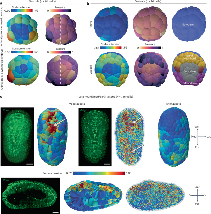
Fig. 5. Spatiotemporal patterning of mechanics in the ascidian embryo P. mammillata.
a, Tension and pressure maps of the animal pole of two 64-cell embryos. Imperfections in the geometric bilateral symmetry of the embryo are reflected by a corresponding asymmetry in the apical tension and pressure of the cell. b, Tension and pressure maps at the animal and vegetal poles of a 76-cell embryo and the corresponding pattern of cell fate in the germ layers. c, Tension maps in late neurula (758 cells) from vegetal, animal and sagittal views. The white arrows indicate regions of higher tension (red) within the embryo. Representative fluorescent microscopy images of myosin II (iMyo) at the vegetal pole and in the sagittal view: 3D reconstruction (top left) or selective plane projection of ten confocal planes (top middle and bottom left). The orientation of the embryo is given by arrows. Ant., anterior; Pos., posterior; Med., medial; Lat., lateral; D, dorsal and V, ventral. Scale bars, 20 μm.

