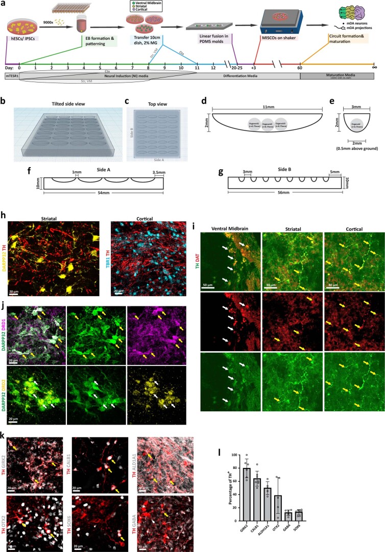
Extended Data Fig. 5. PDMS molds allow fusion of organoids and the observation of dopaminergic innervation.
a, Schematic for the generation of MISCOs. b-c, Tilted side view and top view of PDMS embedding molds for linear fusions. d-e, size considerations for the fusion of three organoids. f-g, Side view of the PDMS embedding mold for MISCO generation. h, Cortical (TBR1+) and striatal (DARPP32+) tissue were innervated by dopaminergic axons (representative images, similar results in n = 8 MISCOs of 2 batches). i, Dopaminergic (TH+) neurons in the VM (white arrow) as well as their axons in striatum and cortex (yellow arrows) of day 90 MISCOs express the mature mDA marker Dopamine Transporter (DAT) on day 90 (representative images, similar results in n = 9/9 MISCOs). j, DARPP32+ neurons in 120-day-old MISCOs in the striatum expressed the striatal subtype markers DRD1 (yellow arrows) and DRD2 (white arrows) (representative images, similar results in n = 6-8 MISCOs of 2-3 batches). k-l, Dopaminergic neurons in MISCOs expressed the dopaminergic subtype markers GIRK2, CALB1, ALDH1A1, OTX2, GABA and SOX6 (n = 6|7|5|5|6|8 MISCOs of 3-4 batches). Yellow arrows: representative double-positive neurons. Data shown as mean ± SD.

