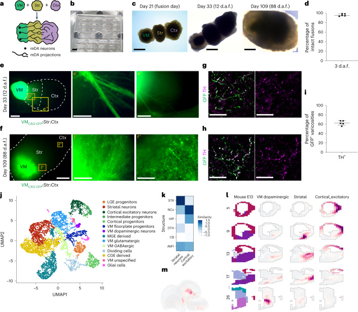
Fig. 2. PDMS mold-assisted spatially arranged fusion of ventral midbrain, striatal and cortical organoids enables the observation of dopaminergic innervation into striatal and cortical tissues.
a, Schematic diagram of the generation of linear anterior–posterior-positioned fused organoids from ventral midbrain (VM), striatal (Str) and cortical (Ctx) organoids. b, PDMS embedding molds with juxtaposed VM–striatal–cortical organoids. c, Triple-fused organoids readily grow together and form a homogeneous tissue. d.a.f., days after fusion. (Representative images; similar results in n = 3–6 organoids per timepoint.) d, Quantification of fusion efficiency of five batches and a total of 365 fusions. A mean of 96% (±2.4% s.d.) of fusions remained intact after the fusion procedure across multiple batches. e, 12 days after fusion (day 33 of organoid age), the first neurite outgrowth can be observed in CtxWT-StrWT-VMCAG-GFP-labeled MISCOs (representative images, similar results in n = 6 of 6 organoids of two batches). Orange and yellow boxes: magnified view in eʹ and eʹʹ. f, On day 109 the striatal (fʹ) and cortical (fʹʹ) tissue are strongly innervated by the VM tissue (representative images; similar results in n = 3 of 3 organoids). Orange and yellow boxes: magnified view in fʹ and fʹʹ. g,h, GFP-positive neurites in cortical (g) and striatal (h) tissue were broadly double positive for the dopaminergic marker TH on day 90 (representative images; similar results in n = 3 organoids per region in cryosectioned tissue, 4–5 organoids of an independent batch in 3D tissue-cleared batch). i, Fraction of dopaminergic varicosities: 63% of all GFP-positive axons in the forebrain were of dopaminergic identity (n = 5 organoids; s.d., 4.9%). j, scRNA-seq of day 60 MISCOs showing that all major populations of the dopaminergic circuit (mDA, striatal and cortical neurons) were present, together with clusters of cortical and LGE progenitors, MGE- and CGE-derived cells and VM GABAergic and glutamatergic neurons (n = 3 pooled MISCOs of one batch). k, Correlation of the cortical excitatory neuron and striatal neuron cluster with the BrainSpan dataset of the developing human brain (postconception weeks 20–25). AMY, amygdala; CB, cerebellum; DTH, dorsal thalamus; HIP, hippocampus; NCx, neocortex; STR, striatum. l, VoxHunt spatial similarity mapping of the cortical excitatory neuron, striatal and VM dopaminergic neuronal clusters onto E13.5 Allen Developing Mouse Brain Atlas data with sections colored by scaled expression similarity scores. m, VoxHunt spatial similarity mapping of the VM dopaminergic cluster onto E13.5 Allen Developing Mouse Brain Atlas data (sagital), colored by scaled expression similarity scores. Data given as mean ± s.d. Scale bars: c (right),f, 1 mm; b,c (left, middle), e, 500 µm; eʹʹ, 250 µm; fʹ, 100 µm; eʹ,fʹʹ, 50 µm; g,h, 20 µm.

