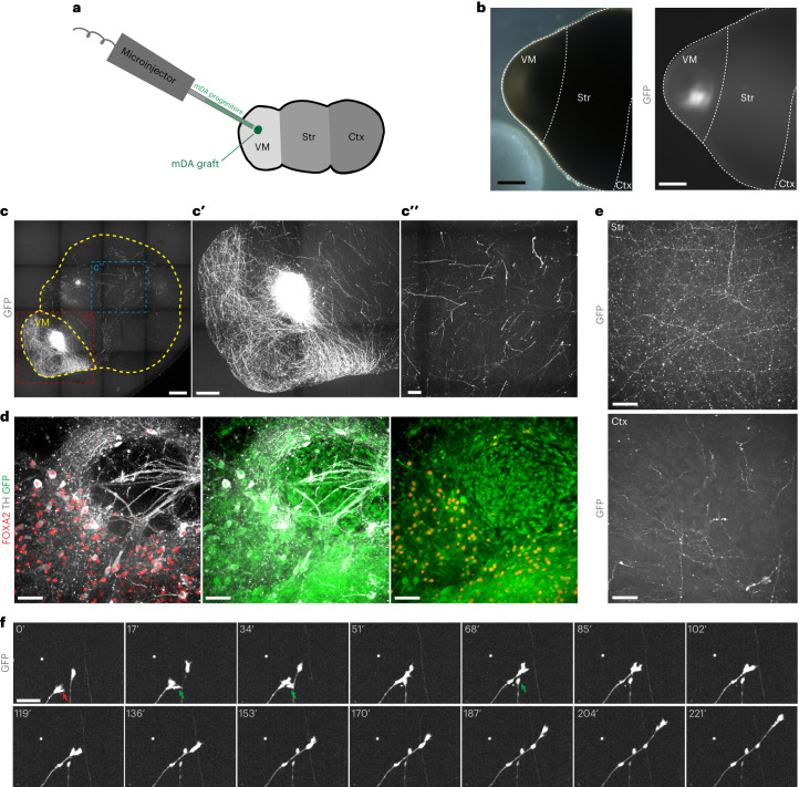
Fig. 5. Injection of high purity ventral midbrain progenitors for Parkinson’s disease cell therapy enables study of graft innervation.
a, Schematic diagram of injection of dopaminergic progenitors into MISCOs. A total of 40,000 cells in a 200 nl volume were injected into the VM part of MISCOs. b, One month after injection, GFP-positive cells were readily visible at the injection site (representative image; similar results in n = 5 of 5 organoids). Left: brightfield of the VM side of a MISCO. Right: same position with GFP fluorescence to visualize injected GFP+ cells. c, Surface recordings 20 days after injection demonstrate graft innervation from VM (cʹ) into striatal and cortical tissues (cʹʹ) (representative images; similar results in n = 3 of 3 organoids). d, 2Eci recording of a graft in VM immunolabeled for GFP, FOXA2 and TH. e, Striatal tissue (top) had denser innervation of grafted cells than cortical tissue (bottom) in 155-day-old organoids. f, Live imaging of MISCOs with VM graft with axon outgrowth into the forebrain tissue. Growth cone (red arrow) and axon–axon interactions (green arrows) were readily observable. Scale bars: b,c, 500 µm; cʹ, 250 µm; cʹʹ, 100 µm; e, 50 µm; d,f, 20 µm.

