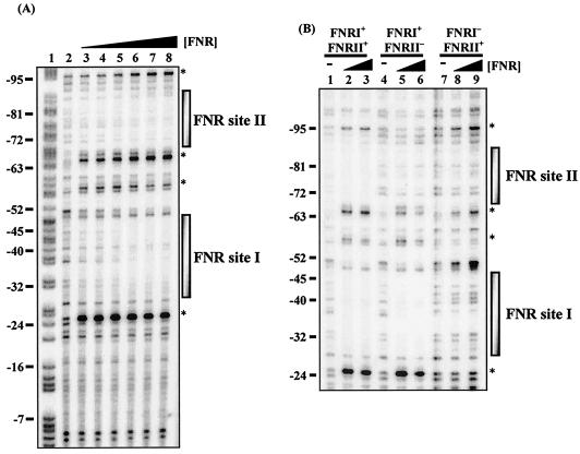
FIG. 4.
DNase I footprint analysis with E. coli FNR for the hyn regulatory region. (A) End-labeled pHYNP3 AatII-HindIII fragment (containing the sequence from position −113 to position 10) incubated with different concentrations of the E. coli Ala154 FNR protein and subjected to DNase I footprinting. The concentrations of FNR in the reaction mixtures were as follows: lane 2, no protein; lanes 3 to 8, 0.125, 0.25, 0.5, 1, 2, and 3 μM, respectively. (B) DNase I footprint analysis for end-labeled pHYNP3 (FNRI+/FNRII+), pHYNP5 (FNRI+/FNRII−), and pHYNP6 (FNRI−/FNRII+) AatII-HindIII fragments. DNA was incubated with no protein (lanes 1, 4, and 7), 1 μM FNR (lanes 2, 5, and 8), and 3 μM FNR (lanes 3, 6, and 9). The gel was calibrated with a Maxam-Gilbert G+A sequencing reaction for the labeled fragment (lane 1), and selected positions are indicated on the left. The shaded boxes indicate the extent of protection afforded by Ala154 FNR binding. The asterisks indicate hypersensitive sites.

