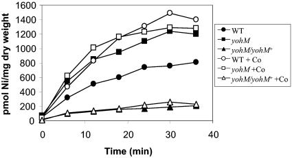
FIG. 3.
63Ni uptake. MC4100 (wt) (circles), ARY023 (yohM::uidA) (squares), and ARY023/pAR020 (yohM::uidA/yohM+) (triangles) cells were grown in Luria-Bertani medium in the presence of 0.5 mM NiCl2 at 37°C under aerobic conditions until mid-log phase (OD600 = 0.6), harvested, and then washed once with buffer consisting of 66 mM KH2PO4-K2HPO4 (pH 6.8) and 0.4% glucose. Cells were resuspended in the same buffer with a fivefold concentration factor. The uptake assay was performed in the presence of 5 μM 63NiCl2 (filled symbols) or 5 μM 63NiCl2 and 50 μM CoCl2 (open symbols). Aliquots (100 μl) were filtered at the indicated times. The intracellular concentration of 63Ni per milligram of bacterial dry weight was determined.

