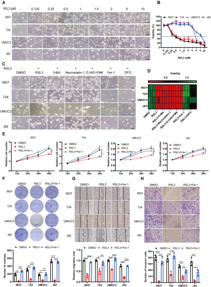
Fig. 1. Ferroptosis induction suppresses proliferation, migration, and invasion of BCa cells in vitro.
A Representative images of BCa cell lines (5637, T24, UMUC3, and J82) treated with various concentrations of RSL3 (0, 0.125, 0.25, 0.5, 1, 2, 5, and 10 μM) for 24 h. Scale bars, 50 μm. B Viability (%) of BCa cell lines treated with RSL3 at different concentrations for 24 h. C Ferroptosis rescue experiment of BCa cell lines treated with RSL3 in combination with various inhibitors: autophagy inhibitor (3-MA, 2 mM), necroptosis inhibitor (Necrostatin-1, 10 μg/ml), apoptosis inhibitor (Z-VAD-FMK, 10 μg/ml), ferroptosis inhibitor (Fer-1, 2 μM; DFO, 100 μM). Scare bar, 50 μm. D Viability (%) of BCa cell lines treated with RSL3 and rescued by various inhibitors for 24 h. E CCK-8 proliferation assay of BCa cells pretreated with RSL3 at suitable concentrations (5637 and T24: 0.125 μM RSL3; UMUC3: 2 μM; J82: 3 μM) that did not affect cell viability. F Colony formation assay. BCa cells were pretreated with RSL3 and grown for ten days. Scale bars, 5 mm. G, H The migratory and invasive ability of BCa cells were assessed by wound healing and transwell invasion assays. BCa cells were pretreated with suitable concentration of RSL3, similar to E). Scale bars, 50 μm. *p < 0.05, **p < 0.01, ***p < 0.001, ****p < 0.0001. The data are represented as mean ± SD of three independent assays.

