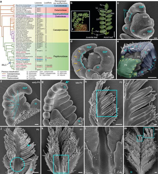
Fig. 1. The ontogeny of compound leaf development in wild type Cicer arietinum (chickpea).
a A new subfamily classification of the Leguminosae is derived from the NCBI taxonomy browser and Azani et al. (2017). The right table summarizes the leaf patterns and leaflet arrangements. The species highlighted in red indicate those that have received relatively extensive study regarding compound leaf development. b Morphology of a representative 6-week-old WT chickpea (cv. CDC Frontiers) (inset), a juvenile leaf (middle) and a mature leaf (right). Scale bars, 1 cm and 2 cm (inset). c–m Morphology of compound leaf primordia at different developmental stages. Images of shoot apical meristem (SAM) with three (c) or four (d) visible leaf primordia showing lateral leaflet (LL) initiation proceeded acropetally (cyan curved arrows) along lateral margins of compound leaf primordia (CLP) and boundaries established between LLs (yellow arrowheads). SAM with five visible leaf primordia (P1-P5) (e) showing trichomes scare at the abaxial surface of the P4 leaf primordium (false-colored in green) but abundant at that of the P5 primordium (blue) with asterisks indicating LL primordia. Adaxial views of leaf primordia showing the distal portion of the CLP differentiated into a terminal leaflet (TL) (cyan arrowhead) primordium during the P4 stage (f, g), the leaflet primordia became folded to adaxial surfaces at the P5 stage (h, i), serration formation (cyan arrowhead) and rachis expansion (dotted cyan circle) at the P6 stage (j), the rachis elongation with trichomes forming on its adaxial surface at the P7 stage (k, l) and trichomes abundant on adaxial surfaces of leaflets (inset) at the P8 stage (m). i, l Close-up views of (h and k). Scale bars, 20 μm in (c, d, f, g, i and l), and 100 μm in (e, h, j, k and m). Similar results were obtained from three biological replicates for each tissue or organ.

