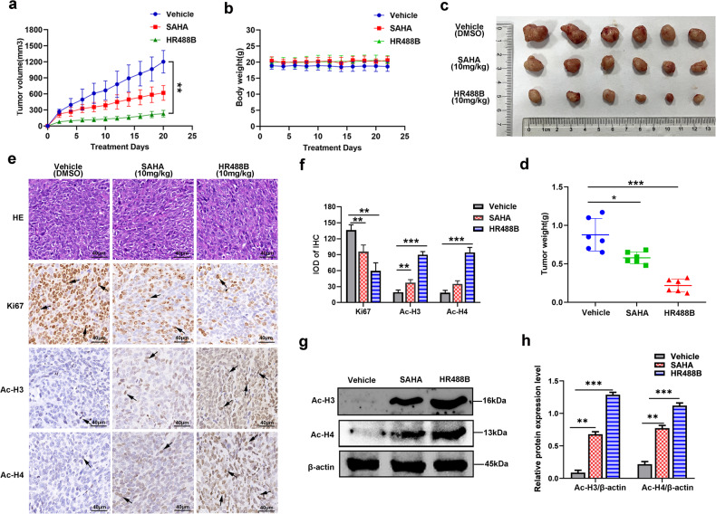
Fig. 3. HR488B suppresses tumor progression in CRC xenograft models.
HCT116 cells (1 × 10⁶) were injected into BALB/c nude mice, and mice were allocated to six groups after 7 days of tumor-cell implantation. a Tumor volume (length × width2 × 0.5) was measured every 2 days and treated with vehicle (5% DMSO in PBS, ip, n = 6), SAHA (10 mg/kg, ip, n = 6) and HR488B (10 mg/kg, ip, n = 6). b The mouse weight was quantified in each group. c The representative stripped images of the tumor entity after being treated with vehicle, SAHA, and HR488B for 2 weeks. d The scatter plot summarized the weight of the tumors. e Representative hematoxylin and eosin (HE) staining of tumor tissues. And immunocytochemical staining for Ki67, Ac-H3, and Ac-H4 expression in tumor tissues from nude mice (Magnification, 400×. Scale bar, 40 μm). f The IHC results were analyzed by Image-Pro Plus 6.0 (n = 5 fields of view). g Western blot analysis of Ac-H3 and Ac-H4 in tumor tissues and β-actin was detected as the endogenous loading control, accordingly. h The statistical result of (g). All data are shown as mean ± SD, two-way ANOVA, *p < 0.05, **p < 0.01, ***p < 0.001.

