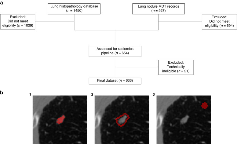
Fig. 1. Recruitment diagram and segmentation labels.
a Study recruitment diagram. Databases of lung histopathology (n = 1450) and lung nodule MDT records (n = 927) were used to identify eligible patients. Following exclusion based on eligibility criteria (n = 1723) and technical limitations of CT images (n = 21), the final internal dataset consisted of 633 patients with 736 nodules. b Cropped, axial plane CT images showing binary segmentation masks (red) for nodule regions. The primary lung nodule was segmented (1) and then expanded by 2 mm isotropically to create a spherical annulus structure (2). An 8 × 8 mm spherical background structure (3) was segmented 15 mm away from the primary lesion.

