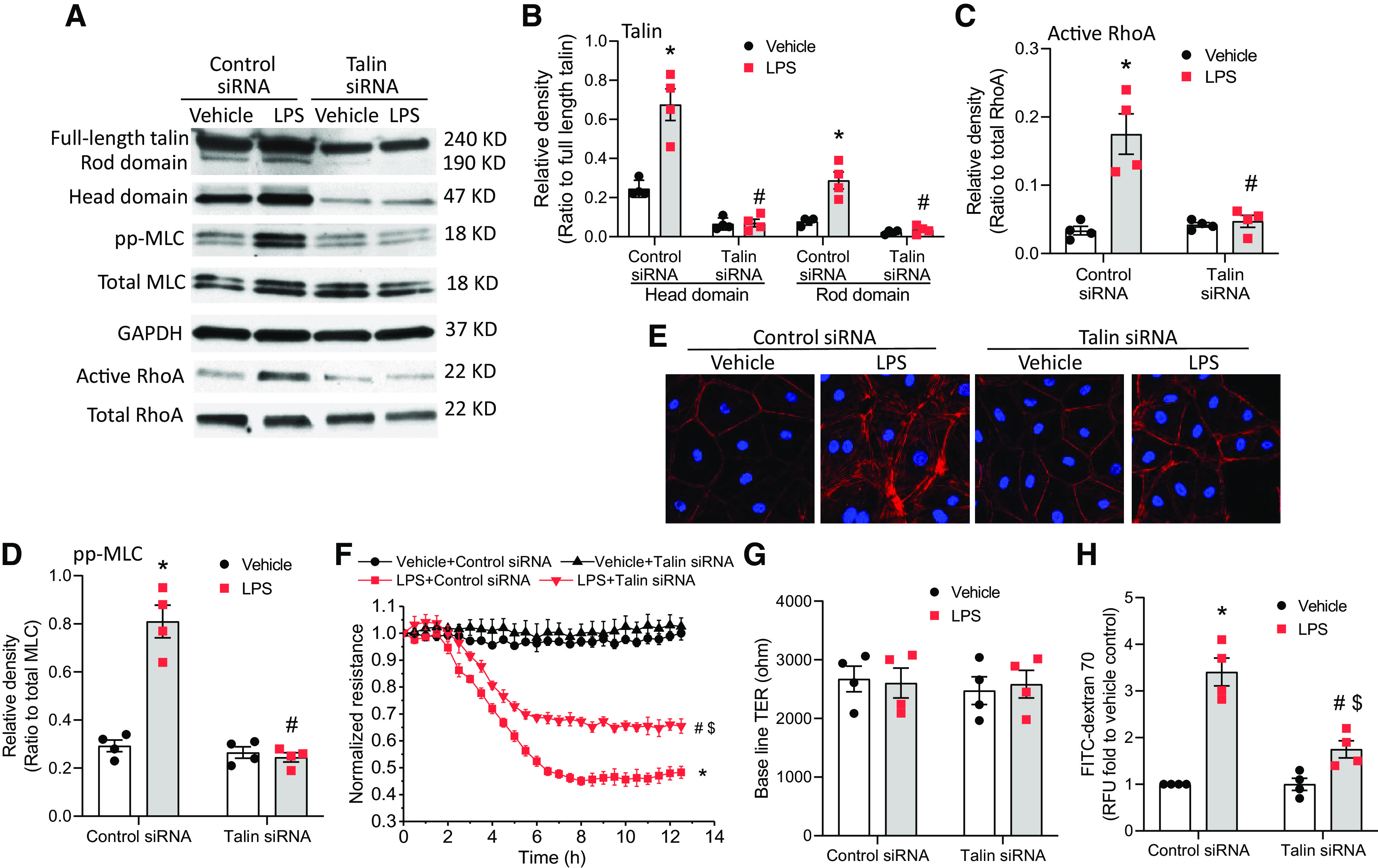
Figure 6.

Knockdown of talin reduces LPS-induced increases in MLC phosphorylation and stress fiber formation in HLMVECs and mitigates LPS-induced lung microvascular endothelial barrier disruption. HLMVECs were transfected with siRNAs against talin or control siRNA. After 72 hours, the cells were incubated with LPS (0.25 μg/ml) for 12 hours, after which the talin head and rod domains, pp-MLC, total MLC, and RhoA activation were measured (A–D); cells were stained using rhodamine phalloidin (E); and HLMVEC monolayer permeability to FITC-dextran 70 (H) was measured and during which the TER value (F and G) was monitored. The blots and images are representative of four independent experiments. Results are expressed as mean ± SE; n = 4. *P < 0.05 versus vehicle + control siRNA; #P < 0.05 versus LPS + control siRNA; $P < 0.05 versus vehicle + talin siRNA.
